Microtissues under the microscope – sample handling with ultrasound
Physicians examine patient cells under a microscope to aid diagnosis, but preparing slides is challenging with small, scarce samples. To prevent loss, a team from CSEM and ETH Zurich developed a method that maximises sample use. In collaboration with ZHAW, it was refined and tested on cancer microtissues.
Looking at cells through the microscope gives physicians an indication of their function or dysfunction. As such, microscopy is the primary tool for physicians to support a medical diagnosis based on patient-derived tissue samples. The process from a tissue sample to a ready-to-analyse microscope slide, however, involves several steps. When the sample is scarce, such as patient-derived cells, every processing step must be optimised to avoid sample loss.
A particular challenge is locating the sample under the microscope. The sample is typically embedded in a so-called microtome block. This is a paraffin block, which is cut into thin slices and then transferred to a microscope slide (see Fig. 1). With a random distribution of the sample in the block, numerous slices must be cut and analysed to obtain an adequate representation of the sample.
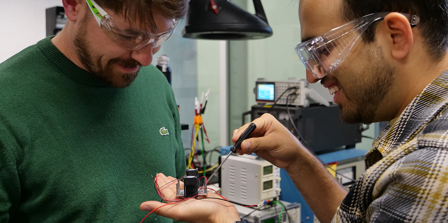
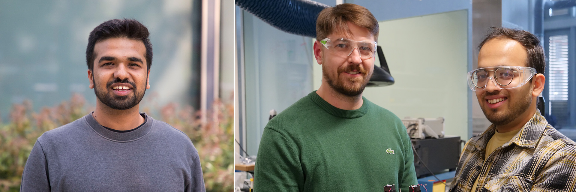
Dhananjay Deshmukh is a tissue engineer and has been in touch with several researchers and industry experts, who seek a user-friendly and efficient process to concentrate the sample in a single plane inside the microtome block. Deshmukh explains: “When slicing such a prepared microtome block in the specified plane, we obtain a maximum of sample on the microscope slide for analysis.”
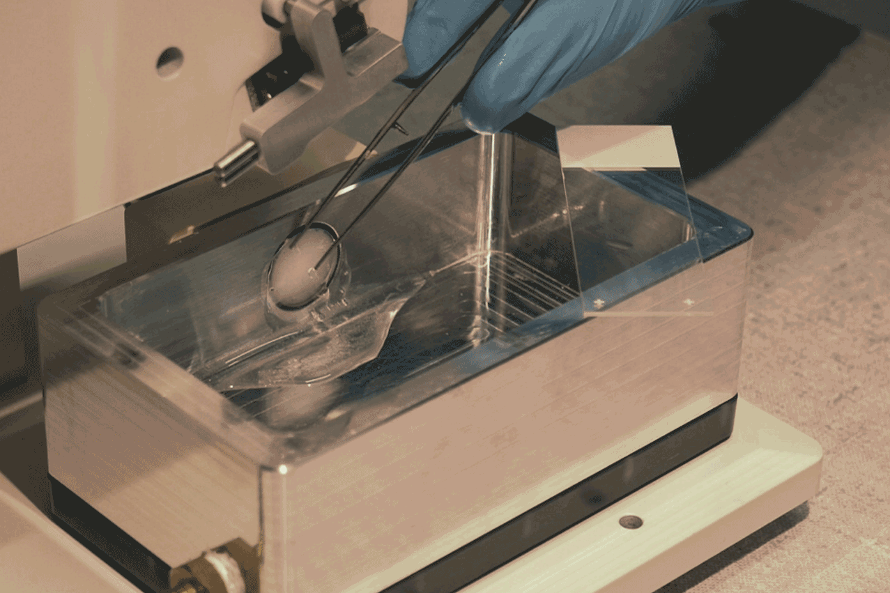
A multidisciplinary research team from CSEM and ETH Zurich has developed an ultrasound-driven mould for the concentration of the sample in a single plane (see Fig. 2). The device creates a standing wave inside an acoustic channel. The microtissues are mixed with a hydrogel and poured into the channel. The sound waves then push the microtissues into the predefined plane. Once the sample is in position, the hydrogel is gelled into a solid block, which is a precursor to the microtome block.
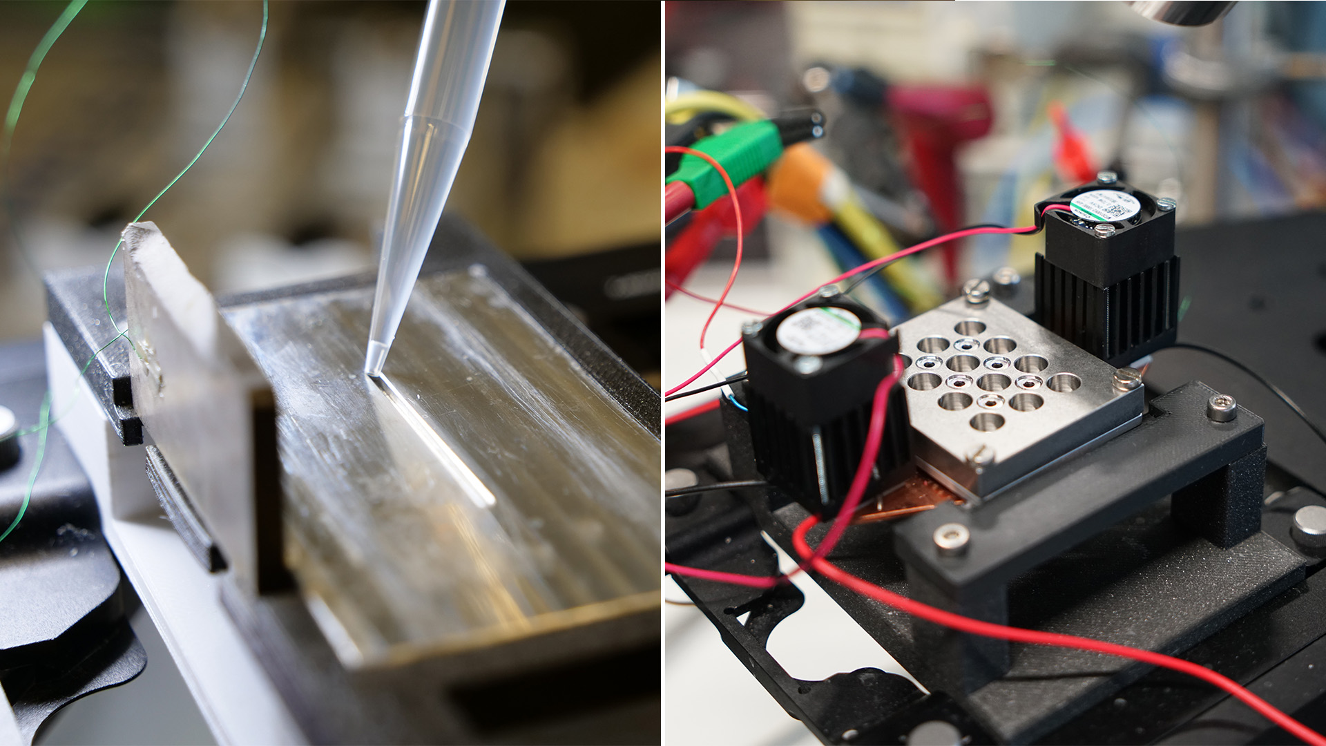
Deshmukh remembers, the first prototype of the ultrasound mould came with several challenges, such as air bubbles in the hydrogel, sticky walls trapping the sample, finding the optimal temperature for pouring, gelling and transferring the hydrogel block. Researchers from CSEM Life Sciences, who are experts in labware, helped with the sample loading mechanism. The combined efforts resulted in a successful proof-of-concept prototype for the ultrasound mould. A joint patent was filed, which is now available for licensing.
Mark Tibbitt, head of the Macromolecular Engineering Laboratory at ETH Zurich, sees a growing demand for tools for microtissue handling: “There is a trend in both industry and academia to replace animal testing with microtissue samples, which are lab-generated mini-organs. A critical roadblock in their implementation is increasing the efficiency of analysis, which our method enables. In collaboration with ZHAW, we have recently demonstrated this with osteosarcoma microtissues.” Gilles Weder, Head of Research & BD Life Sciences at CSEM, confirms: "Following the FDA's recent announcements on alternatives to animal testing, organoid technology continues to gain attention, while micro-histology remains the gold standard for tissue examination."
An invention of ETH Zurich: Dhananjay Deshmukh, Mark Tibbitt, Jürg Dual and CSEM: Emilie Vuile-dit-Bille, Gilles Weder, Sarah Heub
Contact Prof. Mark Tibbitt, Macromolecular Engineering Laboratory
Publication: D. Deshmukh et al., "external page Acoustofluidic patterning for improved microtissue histology", Preprint (2025)
Patent pending, external page PCT/EP2024/060082
Do you want to get more "News for Industry" stories?
external page Follow us on LinkedIn
Are you looking for research partners at ETH Zurich?
Contact ETH Industry Relations